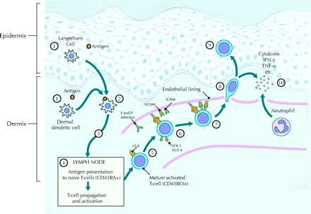
Psoriasis is an autoimmune disorders or a T-helper (Th1) / (Th17) immune dysfunction.T-cell activation, TNFα, and dendritic cells are pathogenic factors stimulated in response to a triggering factor, such as a physical injury, inflammation, bacteria (A self-peptide cross-reacting with streptococcal M protein bacteria-derived superantigens is one of the candidates as streptococcal infections precede 90% of psoriasis type I cases), virus retrovirus (human immunodeficiency virus 1), and human papilloma virus, or withdrawal of corticosteroid medication.
Initially, immature dendritic cells in the epidermis stimulate T-cells from lymph nodes in response to as yet unidentified antigen stimulation . Dendritic cells (DC) act as antigen-presenting cells (APC) to initiate the immunoresponse after stimulation by an as yet unknown signal. Activated DCs migrate to lymphatic tissue and secrete chemokines to attract naive T lymphocytes, which are then activated and differentiated to Th1 and Th17 type cells. Movement of the activated T lymphocytes from the periphery to the skin is elementary for the plaque to develop. the dendritic cells (DC) are the most common type of antigen-presenting cells.
Antigens are presented on the DC surface usually as MHC class II molecules, which can be recognized by T cell receptor of CD4+ T cells. In addition DCs can present antigens on MHC class I molecules, which lead to the activation of CD8+ T cells. DCs also enhance the production of adhesion and co-stimulatory molecules to facilitate its interaction with T cells. The antigens behind the process are not known.
Naive T cells have to go through maturation in lymphatic tissues. CD4+ T cells have a strong affinity to MHC II-peptide complex and they stick together by forming an immunological synapsis. Interaction between ICAM-1, produced by DCs, and LFA-1(lymphocyte function associated antigen 1), from T cells, is one of the most important in order to facilitate the DC-T cell interaction. Naive T cells can mature either to Th1, Th2, Th17 or regulatory T cells and subtype is guided by the selection of a soluble mediator present during the activation of naive T cells. After the activation, T cells express cutaneous lymphocyte-associated antigen (CLA) (cutaneous lymphocyte antigen), which directs T cells to inflamed skin. P- and E-selectins expressed by endothelial cells are also important for T cell skin homing. The invasion is based on tissue-specific ligand receptor interaction and on that account skin homing T cells produce L-selectin, LFA-1 and CLA adhesion molecules.
Resident skin cells also express molecules important in skin homing, including ICAM-1 (intercellular adhesion molecule 1) expressed by epidermal keratinocytes and E- and P-selectins in dermal capillaries. The initial interaction is formed between the L-selectin of T lymphocytes and E- and P-selectins expressed by vascular endothelial cells, after which other ligand-receptor interactions also emerge to continue the inflammation reaction.
Interaction between P- and E- selectins and CLA, as well as other selectin ligands, facilitate leukocytes rolling along the blood vessel wall as they decrease the rolling velocity as T cells recognize chemokines secreted by the endothelial cells, become active and a tight adhesion, facilitated by integrins produced by T cells and their ligands expressed by endothelial cells, is formed between the cells. The most important molecule in skin homing is LFA-1 binding to ICAM-2.
T cells probably enter the endothelial wall through pores formed between endothelial cells in an integrin-dependent process called diapedesis. Both CD4 and CD8 positive T lymphocytes are in psoriasis plaque, CD8+ cells predominantly within the epidermis and CD4+ cells within the dermis. Especially the role of CD4+ cells is critical in the pathogenesis of psoriasis. Other inflammatory cells also take part in pathogenesis but their role is not as well understood. Both macrophages and mast cells produce cytokines including tumor necrosis factor (TNF), interferon γ (IFN-γ) and interleukin 8 (IL-8), which are essential for psoriasis.
Psoriasis is commonly considered to be a Th1 type disease and cytokines of the pathway, including IL-2, IL-12, IFN- γ and TNF-α, are abundantly found in psoriasis plaques Interleukin-2 is a T cell growth factor, which promotes T cell function, as well as stimulates natural killer cell activity, and promotes production of a wide variety of cytokines Interferon γ is an important immuneregulator and also has antiproliferative properties. Furthermore it can induce the expression of ICAM-1 in keratinocytes and endothelial cells and thus influence invasion of skin homing T lymphocytes in psoriatic skin .
Tumor necrosis factor α is produced by keratinocytes and has a focal role in skin inflammatory processes Proinflammatory cytokines IL-6 and IL-8, capable of promoting keratinocyte proliferation, are also expressed abundantly in psoriatic skin . Recent additions to the list of cytokines relevant in psoriasis are IL-12 and IL- 23. In psoriatic skin IL-23 is overproduced by DCs and keratinocytes and promotes Th 17type T cells‘ survival and the production of IL-17 and IL-22, which are also considered to be significant in psoriasis .
Interleukin 17 promotes accumulation of neutrophils, dendritic cells and T cells and also has an impact on barrier function. Interleukin-22 triggers keratinocyte hyperproliferation and down regulates genes associated with keratinocyte differentiation, which are functional aspects relevant for the pathogenesis of psoriasis.
Interleukin-12 participates in several processes relevant for psoriasis including cell proliferation and angiogenesis. It also promotes T lymphocyte activation and differentiation by promoting the Th1 pathway . In addition to the aforementioned cytokines, other cytokines as well as chemokines are found and may have an impact on the pathogenesis of psoriasis.
Toll like receptors (TLRs) of non clonal pattern recognition receptors, which recognize constitutive and conserved molecular patterns shared by many pathogens. TLRs are important in the early recognition of microbial structures. They signal via the transcription factor genes. TLR1, TLR2 and TLR5 were shown to be than that in the normal epidermis. These changes differentiation pathway or result from the effects of proinflammatory cytokines such as TNF-α and IFN-γ present in psoriatic lesion .


0 التعليقات:
إرسال تعليق